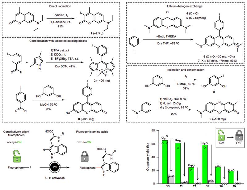
Fluorogenic probes that light up only upon target engagement promise cleaner, faster bioassays than always-on dyes, which demand washing steps to achieve adequate signal-to-noise ratios. Fluorogenic amino acids, FgAAs, are especially attractive because they can be woven directly into peptide sequences via standard solid-phase chemistry, placing the switch right at the binding interface. Yet multicolor FgAAs built from modern, high-brightness fluorophore scaffolds have remained out of reach for two compounding reasons: no intramolecular quenching moiety with broad applicability across different fluorescent cores had been identified, and computational tools to predict the operative quenching mechanism were absent. Without both, each new fluorophore color required independent empirical discovery, and pre-formed fluorogenic dyes reported to date suffer from limited brightness that compromises performance in cell experiments.
Researchers in the Mendive-Tapia and Vendrell Groups at the University of Edinburgh and the Liu Group at Nanyang Technological University, published in Nature Chemistry, reasoned that tryptophan, already present in nature as an electron-rich aromatic, could serve as a universal quenching partner if coupled directly to a fluorophore rather than tethered at distance. The team forged C–C bonds between the indole C2 position of tryptophan and iodinated fluorophore precursors via palladium-catalysed C–H activation, producing compact FgAAs in which quencher and fluorophore share a covalent bond, enabling wash-free bioassays. The authors note that this synthetic strategy could in principle be applicable to a broad range of fluorescent structures by accessing their aryl iodinated precursors, a generality supported in part by computational evidence alongside experimental synthesis of key scaffolds.
As proof of concept, the team built fluorogenic peptides targeting human PD-1 immune-checkpoint receptors; to the best of the authors' knowledge, no fluorogenic probe for human PD-1 had previously been reported. Lead peptide 36 bound PD-1 by surface plasmon resonance and enabled wash-free imaging of activated T cells, while a screen of nine immune-targeting drugs identified avasimibe as a candidate modulator of PD-1 expression, a connection the authors describe as previously undescribed. Full photophysical and cellular imaging data are in the original publication.