Multiple myeloma, MM, is a plasma cell malignancy that infiltrates the bone marrow and remains incurable in most patients. Accurate imaging is essential for staging, treatment selection, and monitoring therapeutic response, yet the most widely used PET tracer, [18F]FDG, struggles with myeloma because tumor cells do not always show strong glycolytic activity and diffuse bone marrow involvement can be difficult to distinguish from background. CD38, a transmembrane glycoprotein with both receptor and ectoenzyme functions, is expressed at consistently high levels on myeloma plasma cells and at much lower levels in normal tissue, making it an attractive molecular target for purpose-built imaging agents.
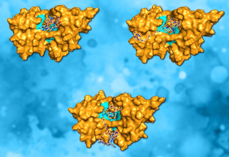
Researchers in the Shokeen Group at Washington University in St. Louis, published in Bioconjugate Chemistry, identified a novel CD38-binding peptide sequence, HAPWFRGGGGS, through phage display screening and built a systematic series of radiolabeled probes around it. The chelator DIAMSAR, whose rigid cage-like sarcophagine structure forms unusually stable complexes with Copper-64, Cu-64, was conjugated directly to the resin-bound peptide during solid-phase synthesis, representing the first reported on-resin DIAMSAR conjugation and eliminating a separate solution-phase coupling step. To address the poor serum stability of the initial l-amino acid construct, Monomer_L, the team synthesized an all-d-amino acid inverso analog, Monomer_D. They then linked two Monomer_D units through a flexible PEG4 spacer to create a dimeric construct, Dimer_D, designed to engage CD38 through multivalent avidity.
The iterative design paid clear dividends at each step. Monomer_L degraded rapidly in mouse serum, retaining only about 45% intact tracer at two hours, whereas Monomer_D held above 92% integrity at four hours, confirming that d-amino acid substitution substantially blocked proteolytic cleavage. Molecular docking predicted tighter binding for Monomer_D than Monomer_L, and cell-binding assays in CD38-expressing MOLP2 human myeloma cells confirmed this, with the d-analog showing significantly higher uptake and a lower Kd of approximately 740 nM compared with 1043 nM for the l-form. Dimerization did not substantially lower the Kd further but nearly doubled the maximum binding capacity, Bmax, from 3024 to 6993 fmol/mg, a hallmark of avidity-driven multivalent engagement. In living animals, the differences were striking: in a disseminated MOLP2 myeloma model, [64Cu]Cu-Dimer_D achieved femoral uptake of 2.93% ID/mL at two hours post-injection, nearly double that of [64Cu]Cu-Monomer_D and more than three times the signal seen in naive controls. In a subcutaneous tumor model, the dimer delivered a tumor-to-muscle ratio exceeding 10, and autoradiography of excised tissues confirmed tracer concentration in CD38-positive lesions.
The work establishes a modular blueprint for building CD38-targeted PET probes from a phage display-derived peptide lead: on-resin DIAMSAR conjugation simplifies synthesis, d-amino acid inversion solves metabolic stability, and dimerization converts modest monovalent affinity into robust imaging performance through avidity. Elevated kidney and liver uptake observed with the dimeric construct signal that pharmacokinetic optimization remains a next step, and the authors plan to explore higher-order multimeric designs and evaluate the approach in primary cells and additional MM models. The strategy is expected to generalize to other peptide-based targeting systems where enhanced receptor engagement and favorable clearance kinetics are jointly required.